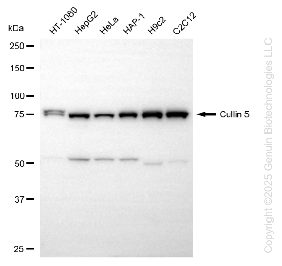
Western blotting analysis using anti-IHH antibody (Cat#5646). Total cell lysates (30 μg) from various cell lines were loaded and separated by SDS-PAGE. The blot was incubated with anti-IHH antibody (Cat#5646, 1:5,000) and HRP-conjugated goat anti-rabbit secondary antibody (Cat#201, 1:20,000) respectively. Image was developed using NaQ™ ECL Substrate Kit (Cat#716).
Flow cytometric analysis of IHH expression in C2C12 cells using anti-IHH antibody (Cat#5646, 1:2,000). Green, isotype control; red, IHH.
IHH; Indian Hedgehog Signaling Molecule; HHG2; BDA1; Indian Hedgehog Protein; Indian Hedgehog (Drosophila) Homolog; Indian Hedgehog Homolog; EC 3.1.-.-; HHG-2
Western blotting analysis using anti-IHH antibody (Cat#5646). Total cell lysates (30 μg) from various cell lines were loaded and separated by SDS-PAGE. The blot was incubated with anti-IHH antibody (Cat#5646, 1:5,000) and HRP-conjugated goat anti-rabbit secondary antibody (Cat#201, 1:20,000) respectively. Image was developed using NaQ™ ECL Substrate Kit (Cat#716).
Flow cytometric analysis of IHH expression in C2C12 cells using anti-IHH antibody (Cat#5646, 1:2,000). Green, isotype control; red, IHH.